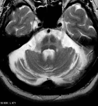
Hot Cross Buns Sign
Axial MRI
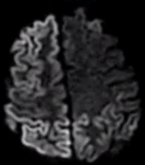
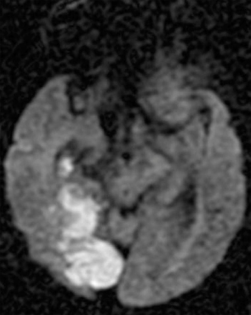
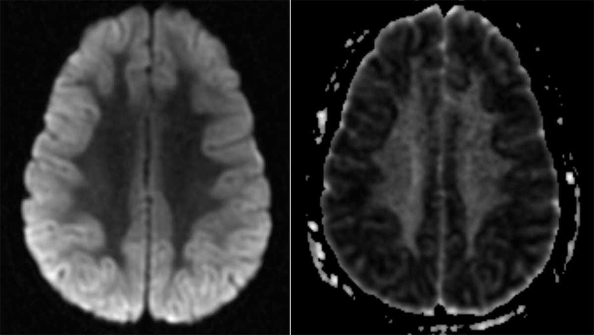
Cortical Ribboning of CJD
DWI Axial MRI
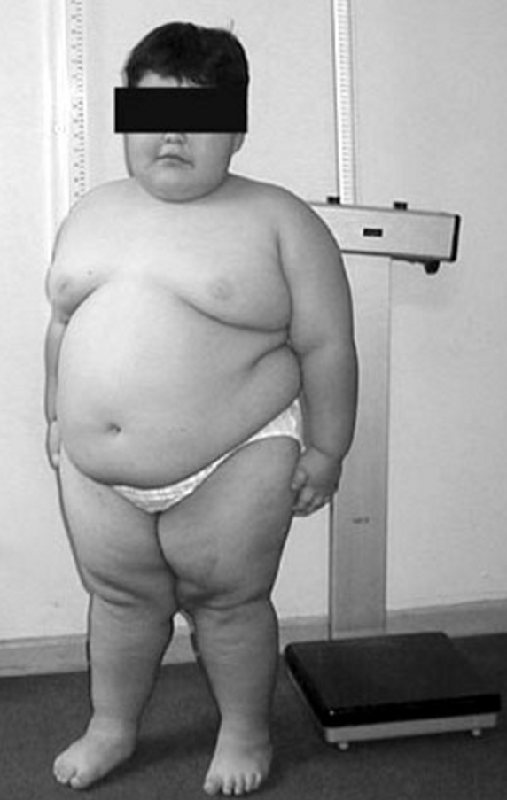
Prader Willi syndrome
8-year-old child with Prader Willi syndrome
Attestation: Fanny Cortés M1, M. Angélica Alliende R1,a, Andrés Barrios R1,2, Bianca Curotto L1,b, Lorena Santa María V1,c, Ximena Barraza O3, Ledia Troncoso A2, Cecilia Mellado S4,6, Rosa Pardo V, CC BY 4.0 , via Wikimedia Commons
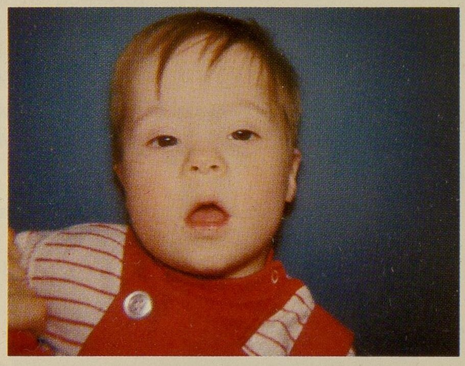
Trisomy 21
Child with classic facial characteristics of Trisomy 21
Attestation: Sydney S. Gellis and Murray Feingold, Public domain, via Wikimedia Commons
Child with Fragile X syndrome
Note the characteristic long ears and face in a patient with Fragile X syndrome
Attribution: Peter Saxon, CC BY-SA 4.0, via Wikimedia Commons
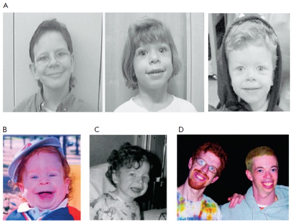
Williams syndrome
Distinctive facial appearance of persons with WS (A). Young child with WS at the age of 15 months (B) and 3 years (C). Note subtle characteristic facial features, including wide mouth, chubby cheeks, long philtrum, small nose, and delicate chin. The same patient is shown in Figs. 1B, 1C, and 1D (left; 21 years); another individual with WS aged 28 years is shown in Fig. 1D (right).
Attestation: E. A. Nikitina, A. V. Medvedeva, G. A. Zakharov, and E. V. Savvateeva-Popova, 2014 Park-media Ltd, CC BY 3.0 , via Wikimedia Commons
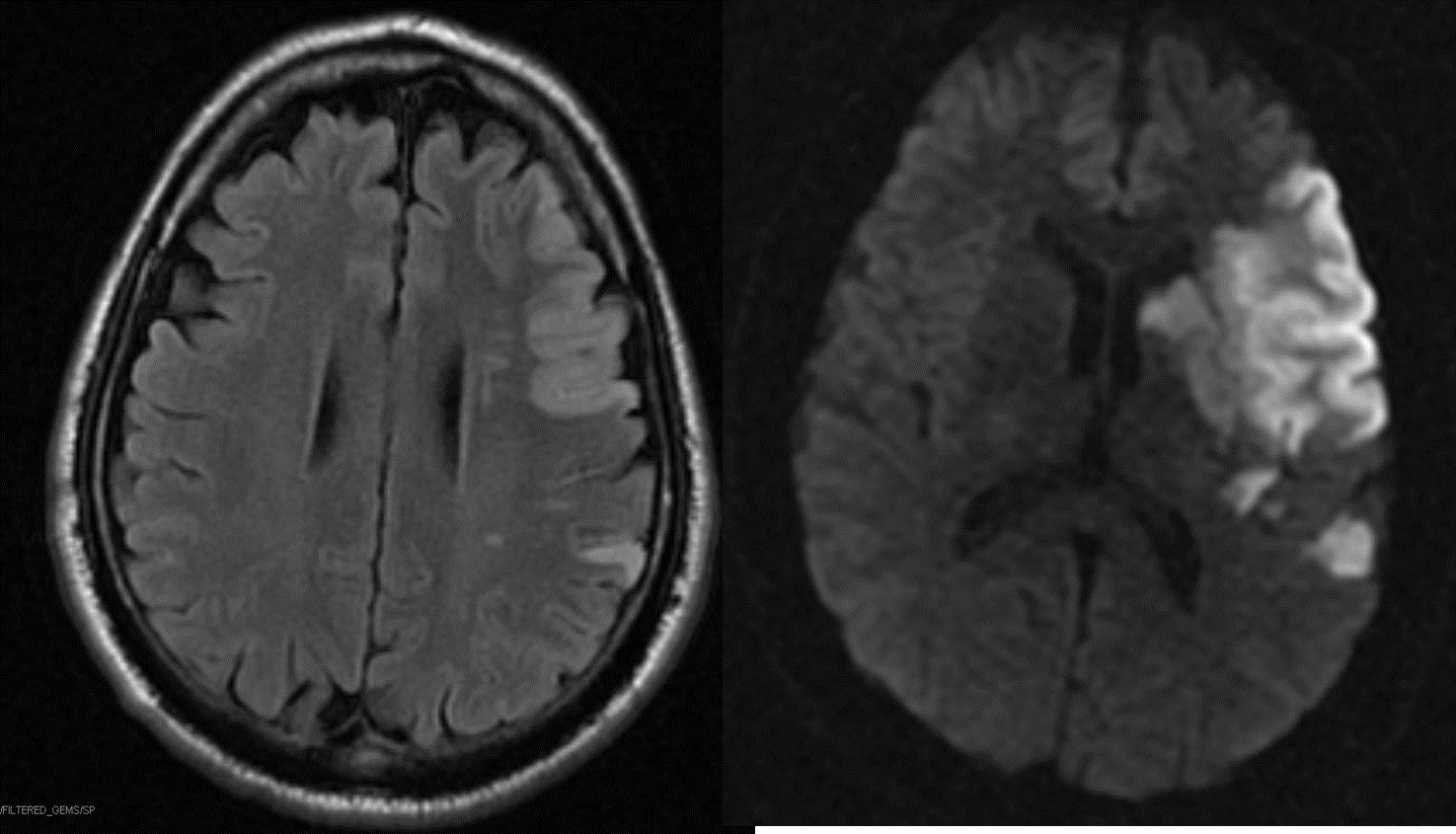
L MCA stroke
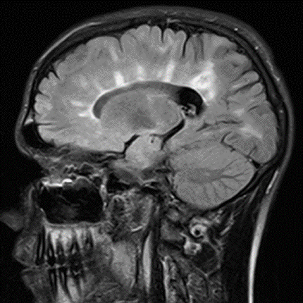
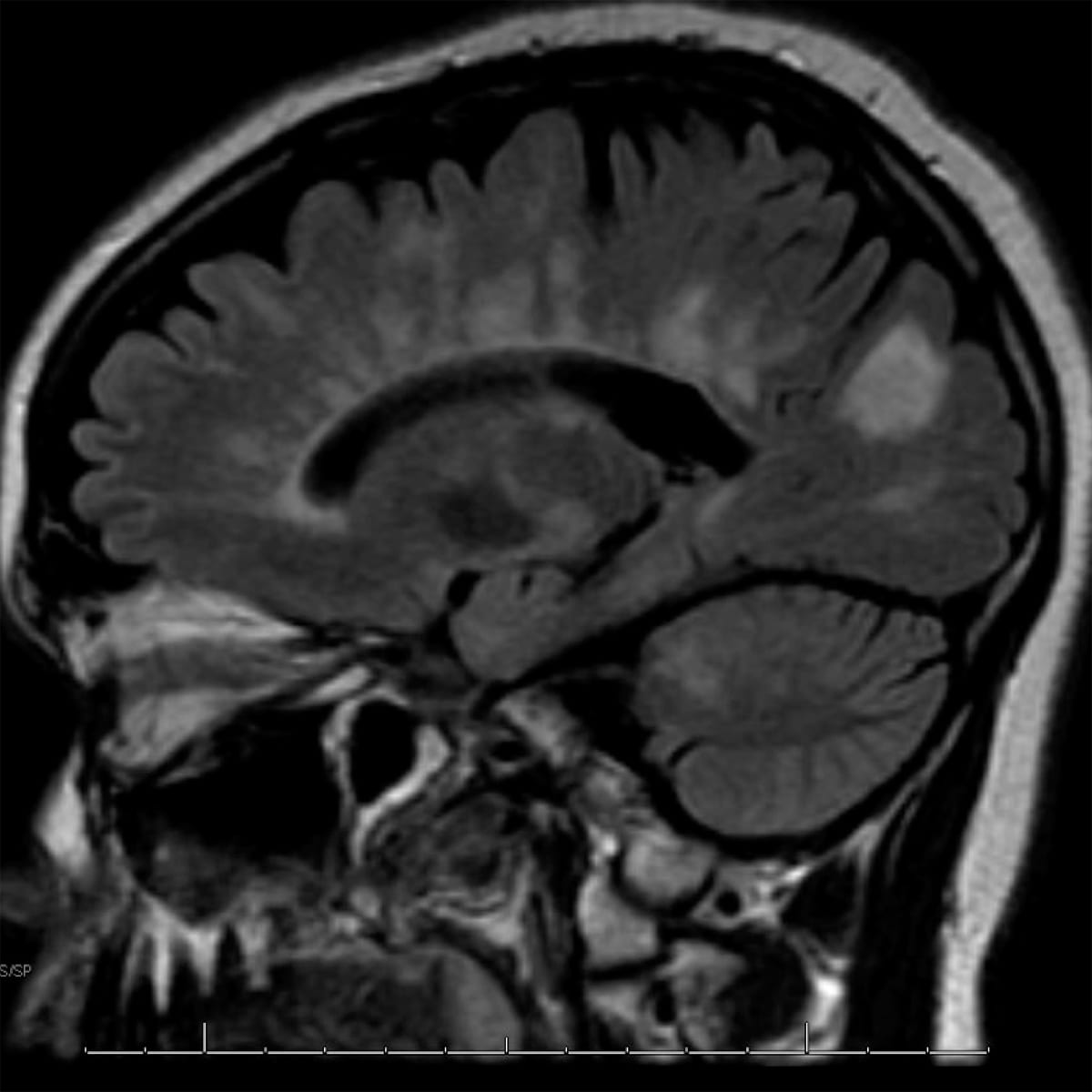
dawsons fingers
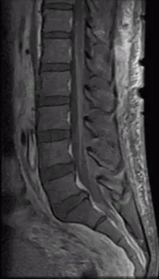
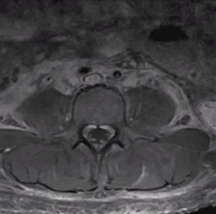
Gullian Barre with enhancement of the nerve roots
MRI lumbar spine with contrast showing contrast enhancement of the nerve roots
Guillan-barre-with-enhancement-of-the-nerve-roots
CJD cortical ribbening DWI
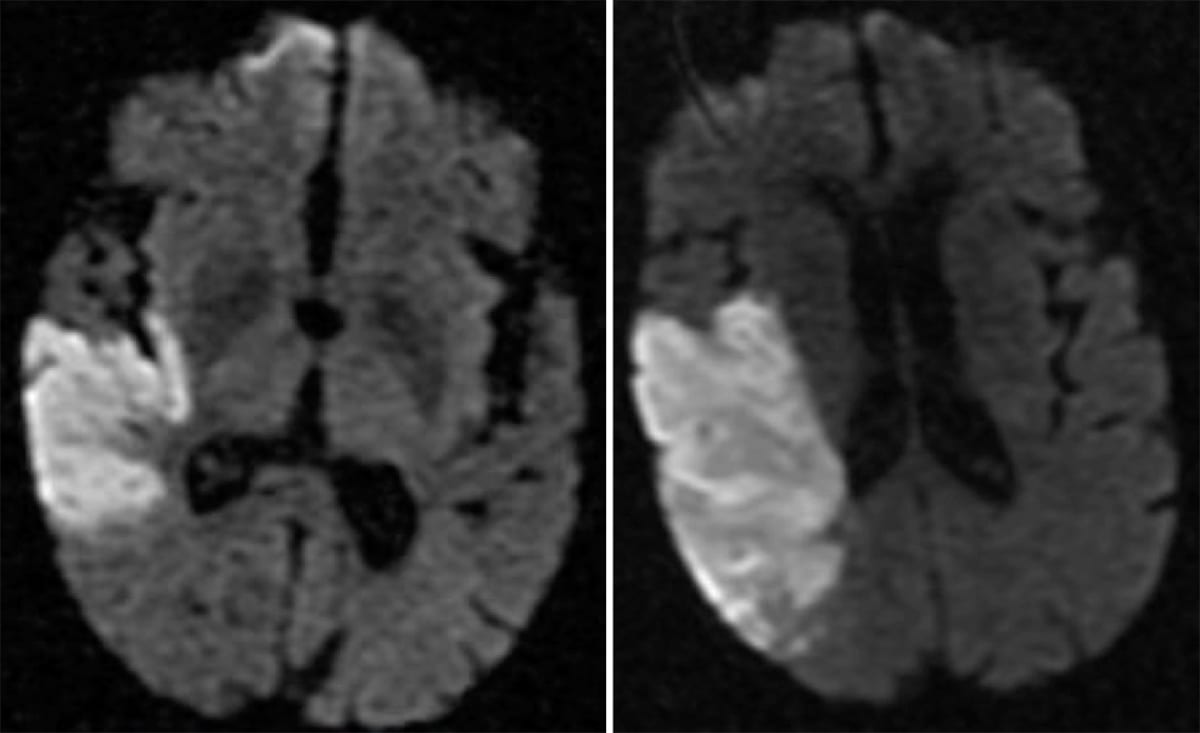
r_mca_acute_infarct_dwi
Right MCA territory infarct, MRI brain, DWI sequence, axial cut
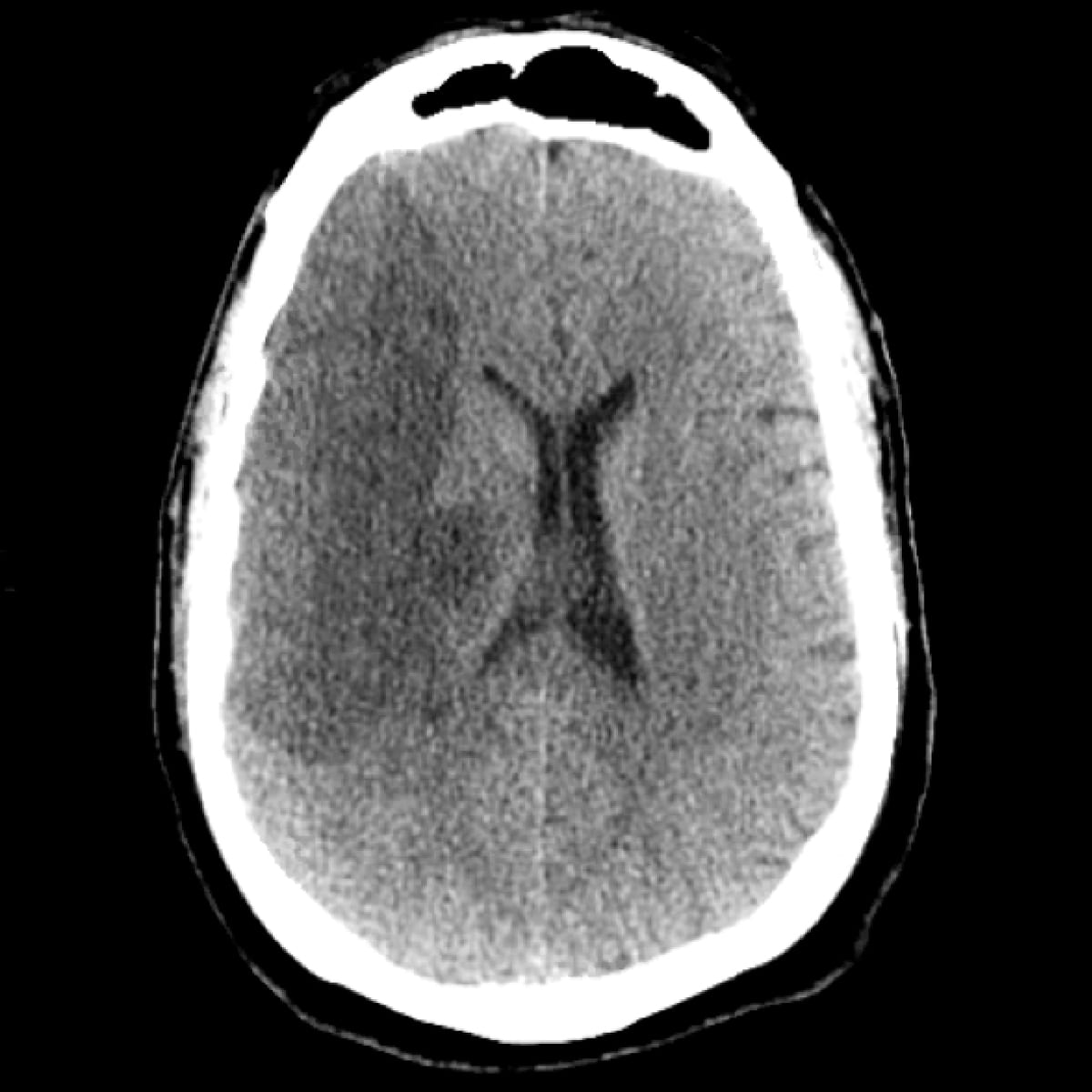
r_mca_stroke_chronic_cth
Chronic right MCA territory stroke, CT scan, axial cut
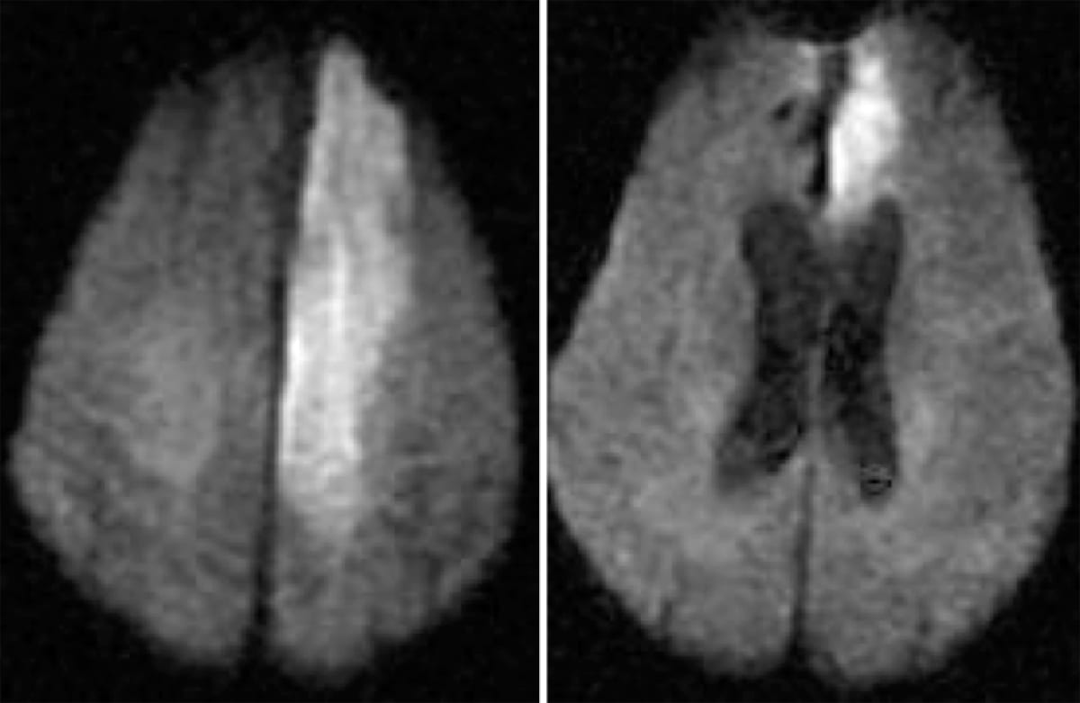
l_aca_infarct_dwi
Acute left ACA territory infarct, MRI Brain, DWI sequence, axial cut
acute_r_pca_stroke_dwi-814x1024
Acute right PCA territory infarct, MRI brain, DWI sequence, axial cut
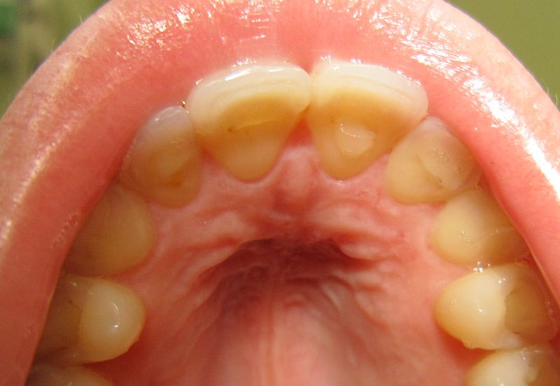
Oral_Manifestation_of_Bulimia.
Bulemia Enamal Loss
Enamel loss of the teeth due to bulimia
chiari_1
Chiari 1 Malformation
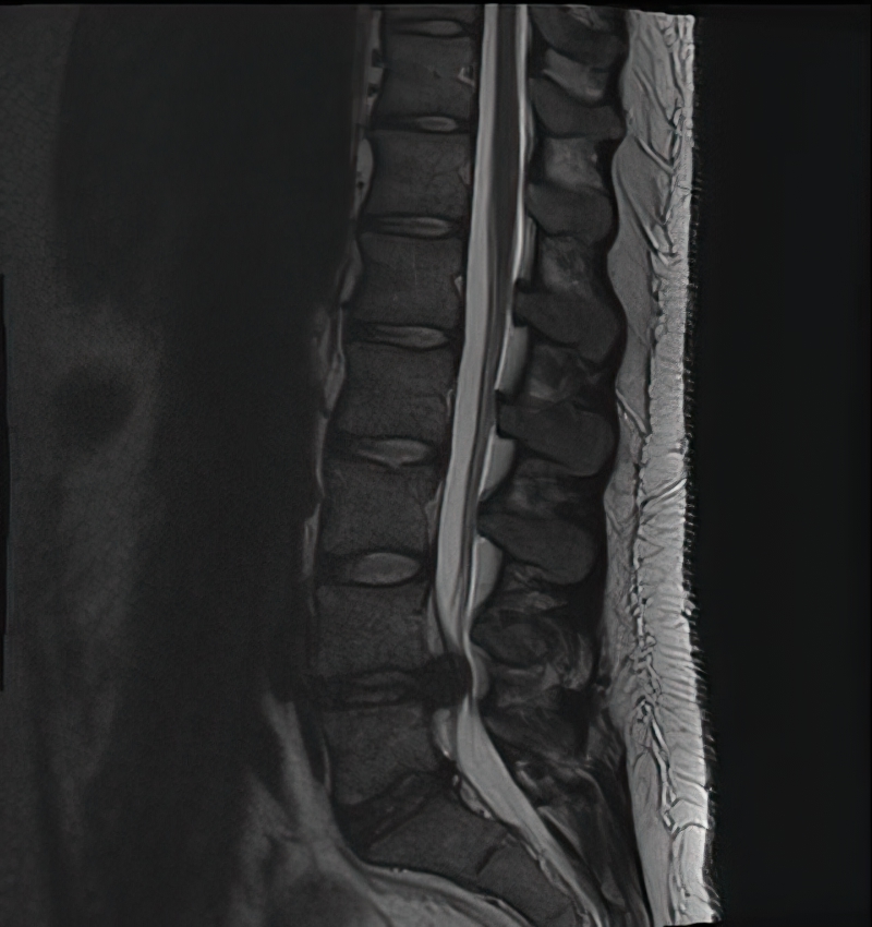
L4-5-disc-herniation
L4-L5 Disc Herniation
chiari_1_holocord_syrinx_flair
Chiari 1 Malformation with Holocord Syrinx
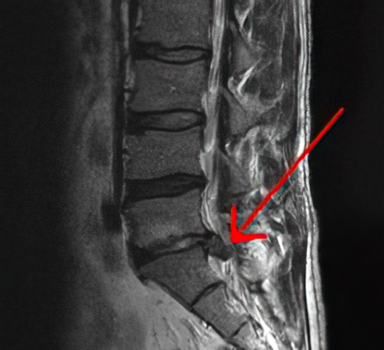
S1-disc-herniation
S1 Disc Herniation
L MCA Occlusion
L MCA Occlusion
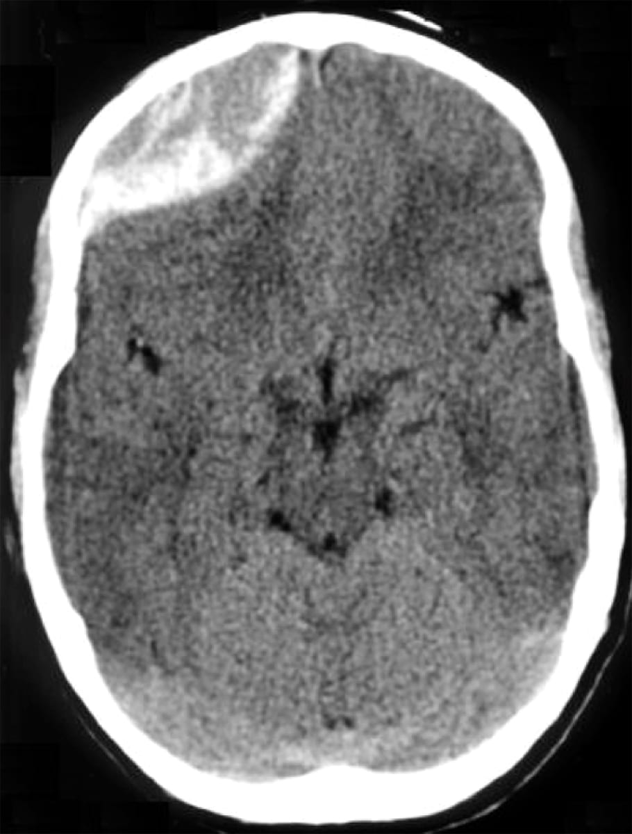
Cerebral Contusion on CT head
Cerebral Contusion on CT head
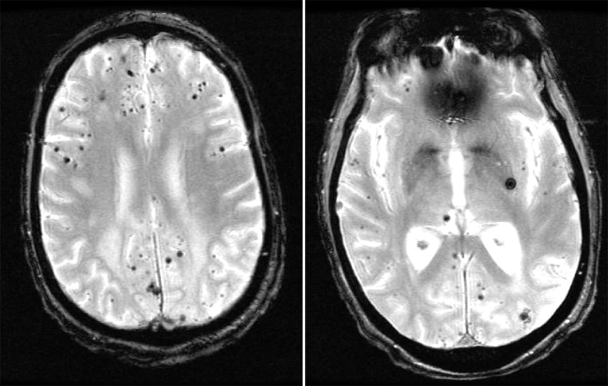
GRE Microbleeds Cerebral Amyloid Angiopathy
Cerebral Amyloid Angiopathy
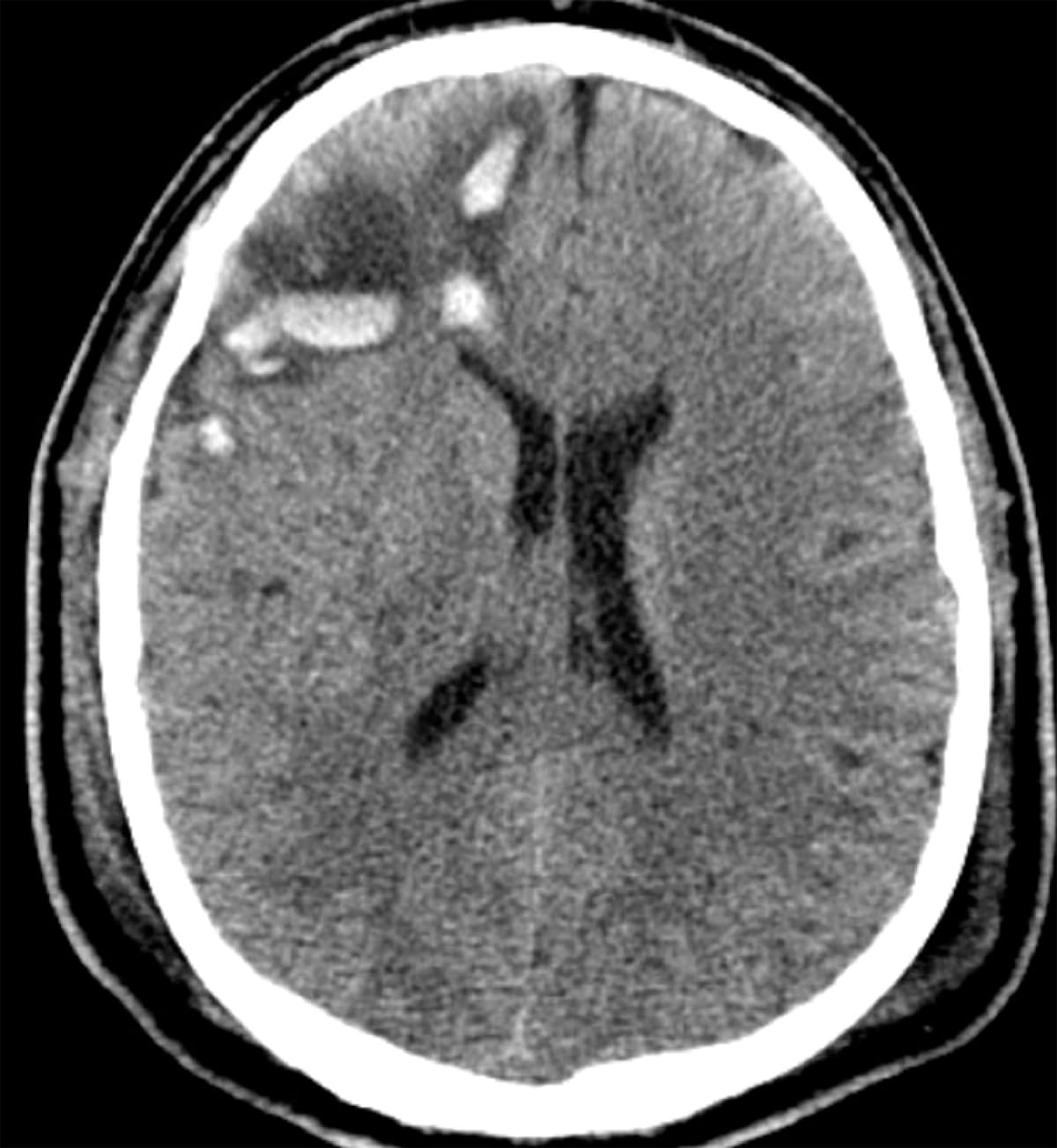
Chronic Left Subdural Hematoma
Chronic Left Subdural Hematoma
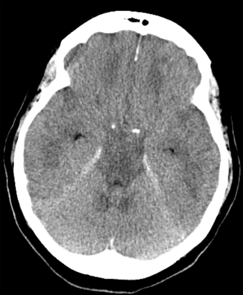
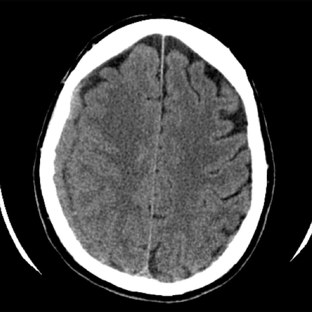
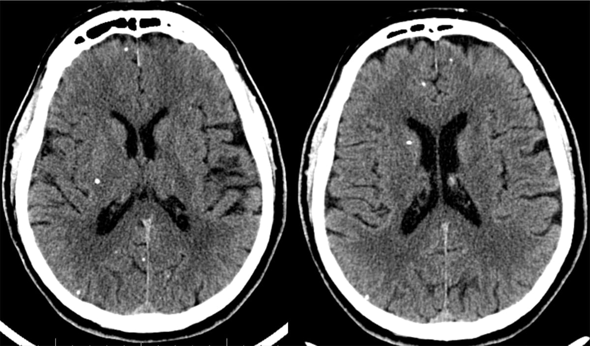
Diffuse Hypoxic Damage on CT after Cardiac
Diffuse Hypoxic Damage after Cardiac Arrest
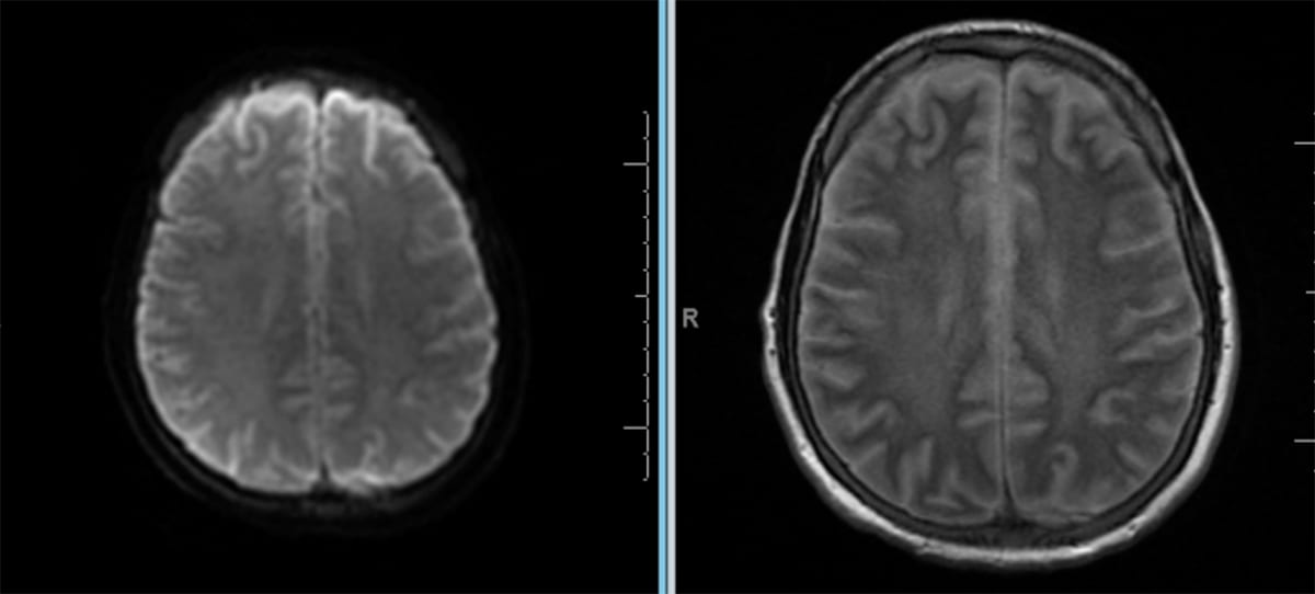
hie_cardiac_arrest_dwi_t2flair_diffuse_hyperintense
Hypoxic Ischemic Encephalopathy
hie_dwi_adc_diffuse_diffusion_restriction
Hypoxic Ischemic Encephalopathy
R Epidural Hematoma CT
Right Epidural Hematoma
Acuter R SDH CT
Acute Right Subdural Hematoma
Dawson's Fingers in Multiple Sclerosis
Dawson's Fingers in Multiple Sclerosis
SAH with vent spread ct 2
Subarachnoid Hemorrhage
Parkinson Substantia Nnigra
Parkinson's Disease
Chiari 1
Chiari 1 Malformation
Chiari 1 Malformation with Holocord Syrinx on MRI
Chiari 1 Malformation with Holocord Syrinx
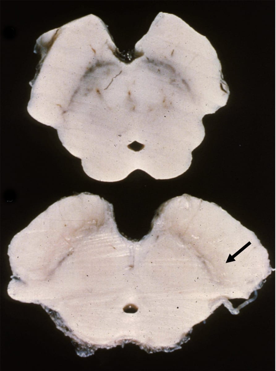
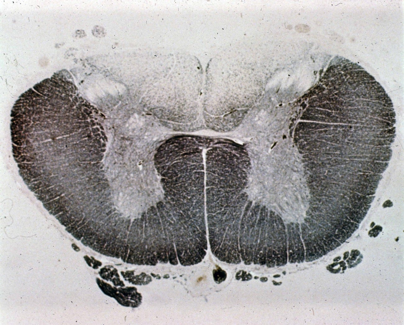
Tabes Dorsalis
Tabes Dorsalis
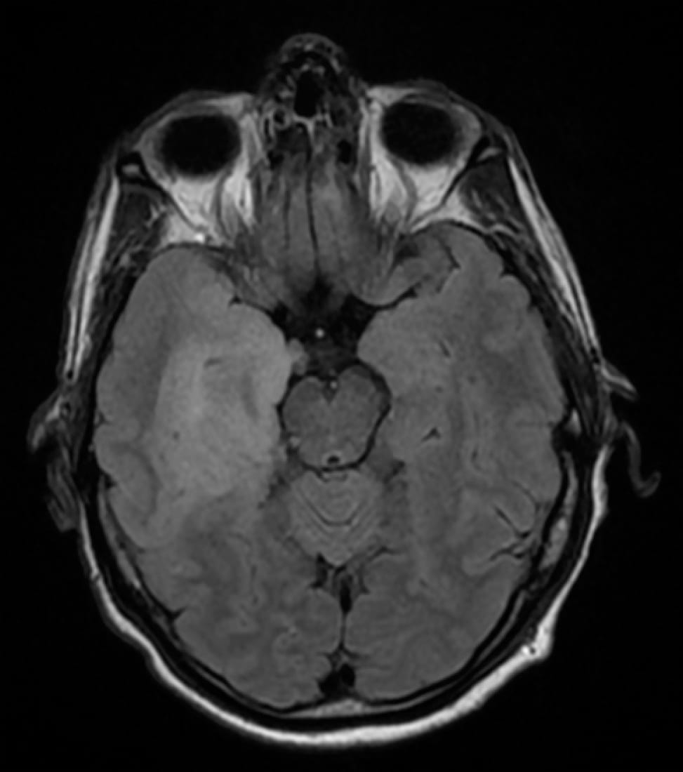
Herpes Encephalitis MRI
HSV Encephalitis: MRI T2 FLAIR sequence with bilateral temporal lobe hyperintensities.
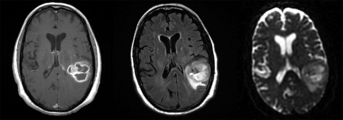
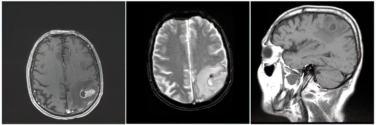
GBM T1 w/ contrast FLAIR ADC
Glioblastoma Multiforme (GBM). Left: Axial MRI, T1 w/ contrast. Middle: T2 FLAIR. Right: ADC.
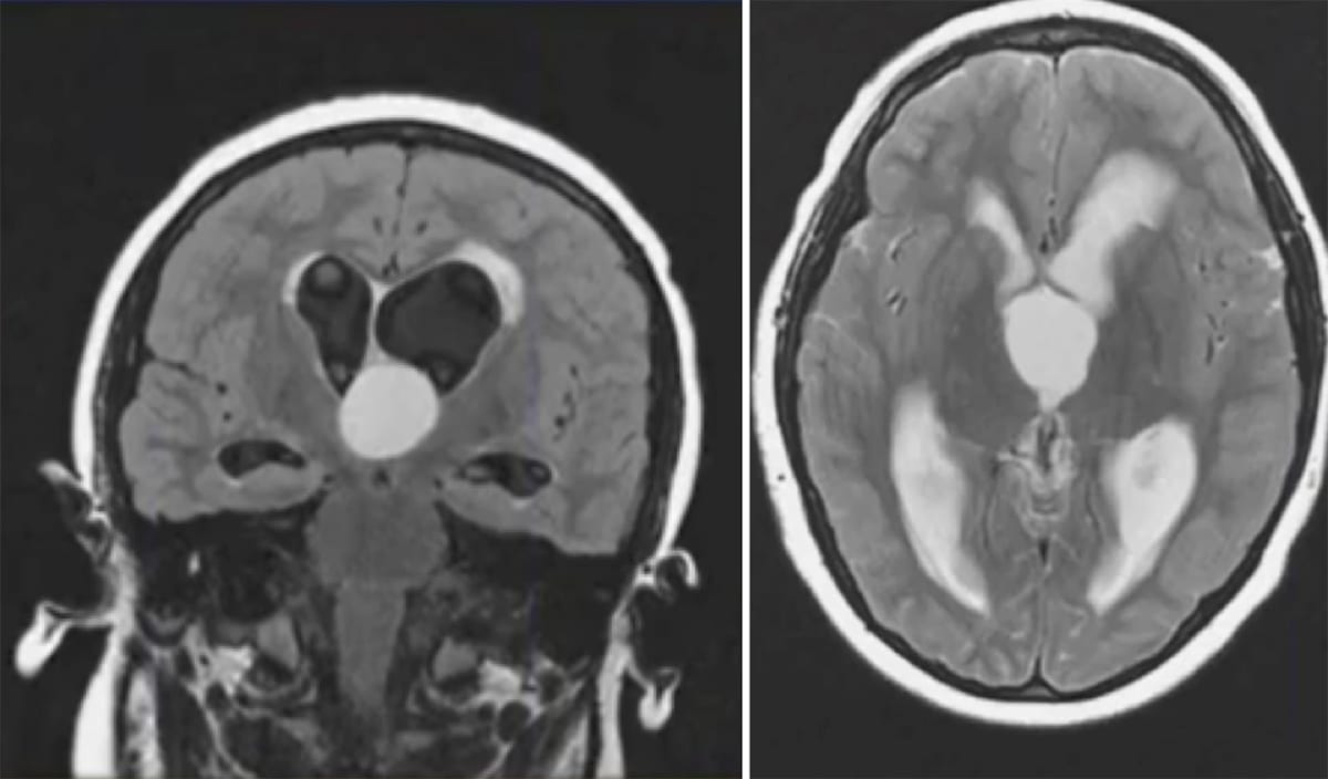
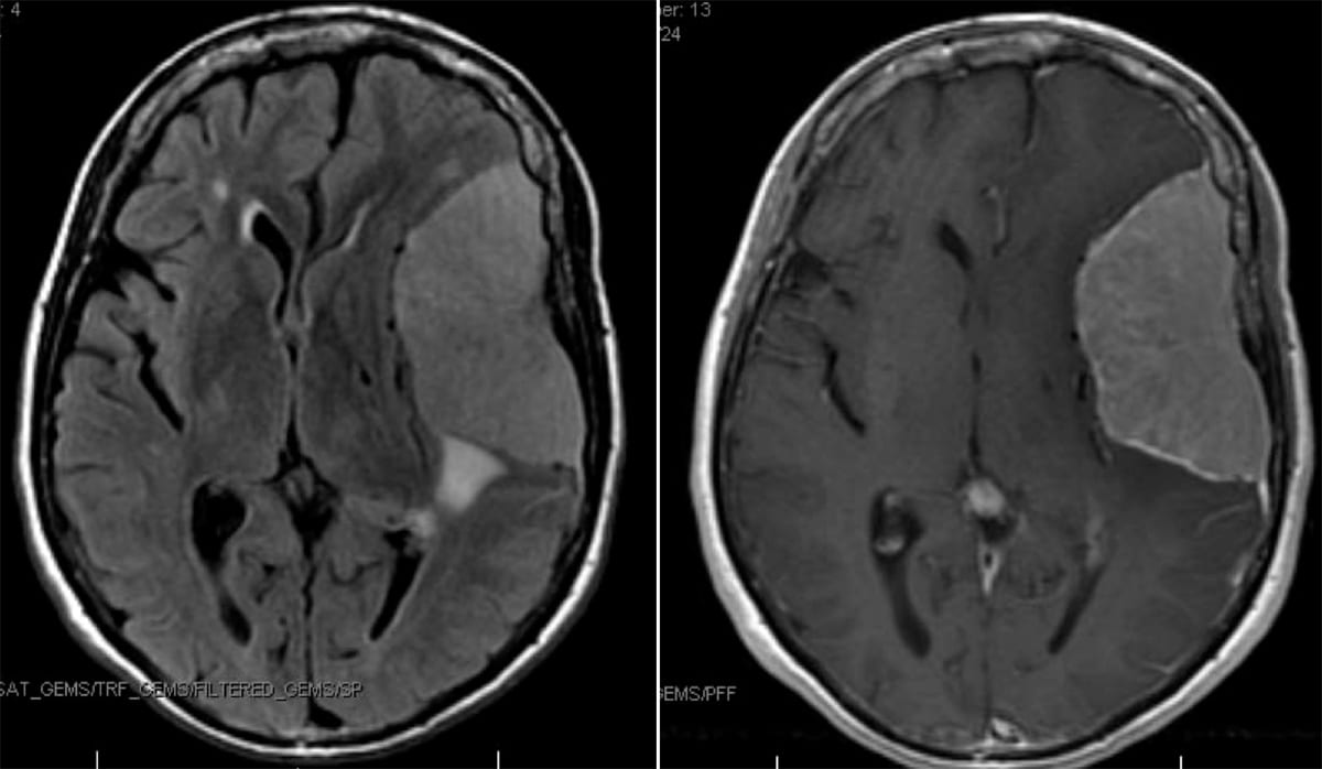
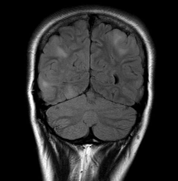
Colloid Cyst T2 FLAIR
Colloid Cyst. Left: Coronal T2 FLAIR MRI. Right: Axial T2 MRI.
Old Cysticercosis CT
Cysticercosis: Axial CT head showing multiple calcified lesions throughout the brain suggestive of a previous infection with cysticercosis.
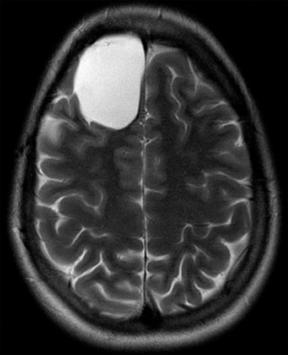
T1 Arachnoid Cyst
Right frontal arachnoid cyst. Axial MRI, T2 sequence.
T2 FLAIR_T1con Enhance Meningioma
Meningioma. Left: Axial MRI, T2 FLAIR. Right: T1 w/ contrast (enhancing).
Toxo T1 Enhanced
Toxoplasmosis
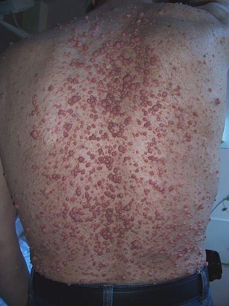
Neurofibromomatosis-type-1-with-cutaneous-neurofibromas
Cutaneous Neurofibromas
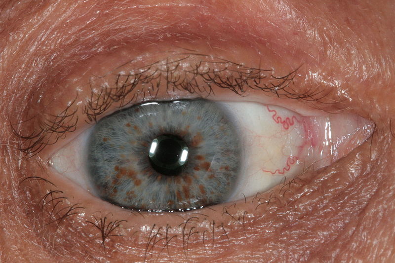
Lisch_nodules-NF-1
Lisch nodules
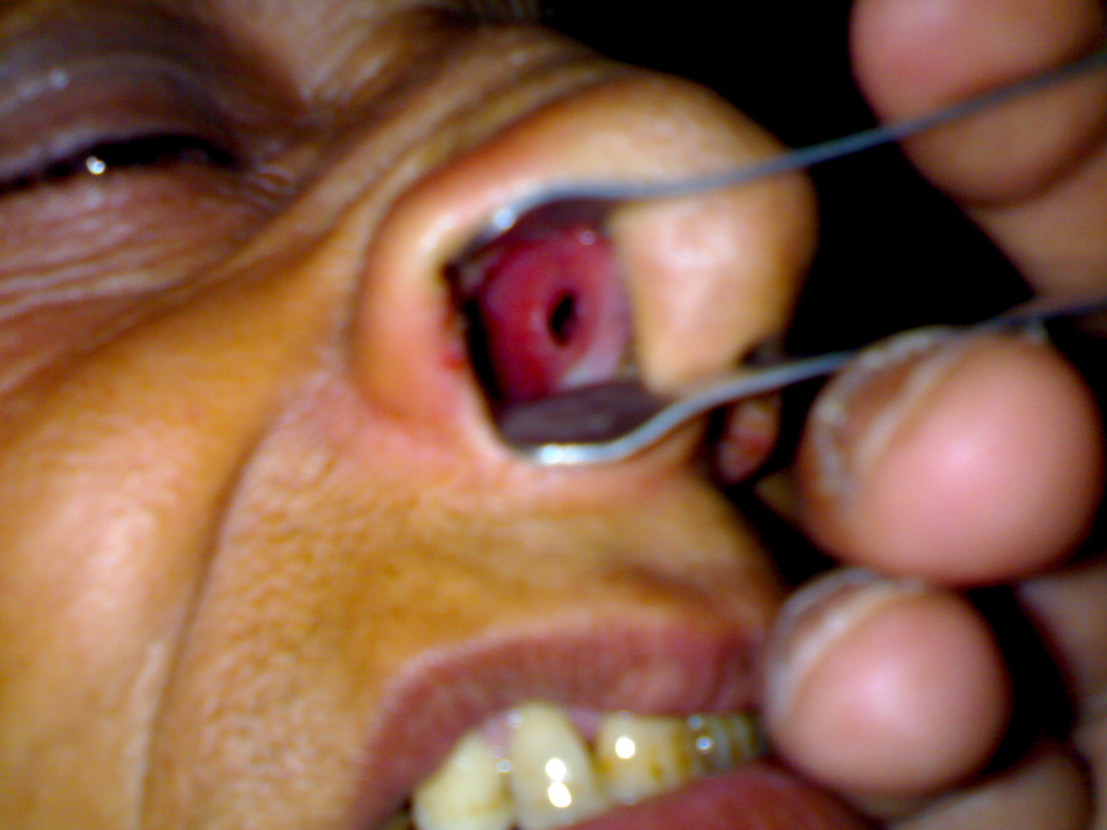
Septal Perforation
Example of a perforated nasal septum.
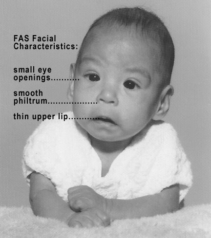
Photo of baby with FAS
Baby with Fetal alcohol syndrome.
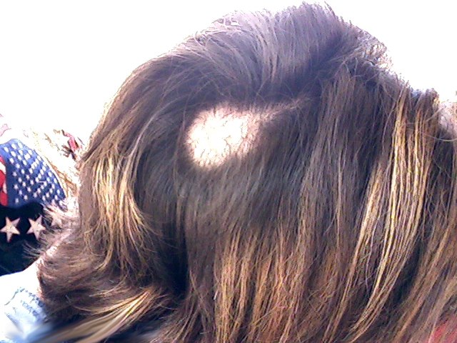
Example of patchy hair loss secondary to trichotillomania
Example of patchy hair loss secondary to trichotillomania
Brain Abscess T2 FLAIR
Cerebral Abscess. Left: T2 FLAIR. Middle: T1 w/ contrast. Right: T1 w/o contrast.
Tuberous sclerosis numerous scattered areas of abnormal T2 signal hyperintensity and cortical thickening consistent with cortical and subcortical tubers
Note numerous scattered areas of abnormal T2 signal hyperintensity and cortical thickening consistent with cortical and subcortical tubers consistent with tuberous sclerosis
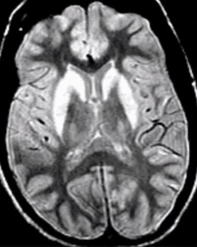
Wilsons disease T2 hyperintensity in putamin and caudate
Axial MRI, T2 sequence, with hyperintensity in the bilateral caudate and putamen
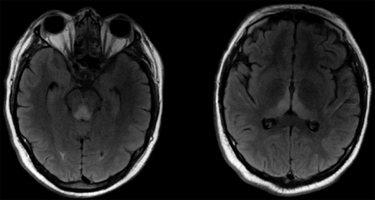
Wernicke's Encephalopathy on Axial T2 FLAIR MRI
Wernicke's Encephalopathy on Axial T2 FLAIR MRI showing hyperintensity of the periaqueductal grey and dorsomedial thalamus
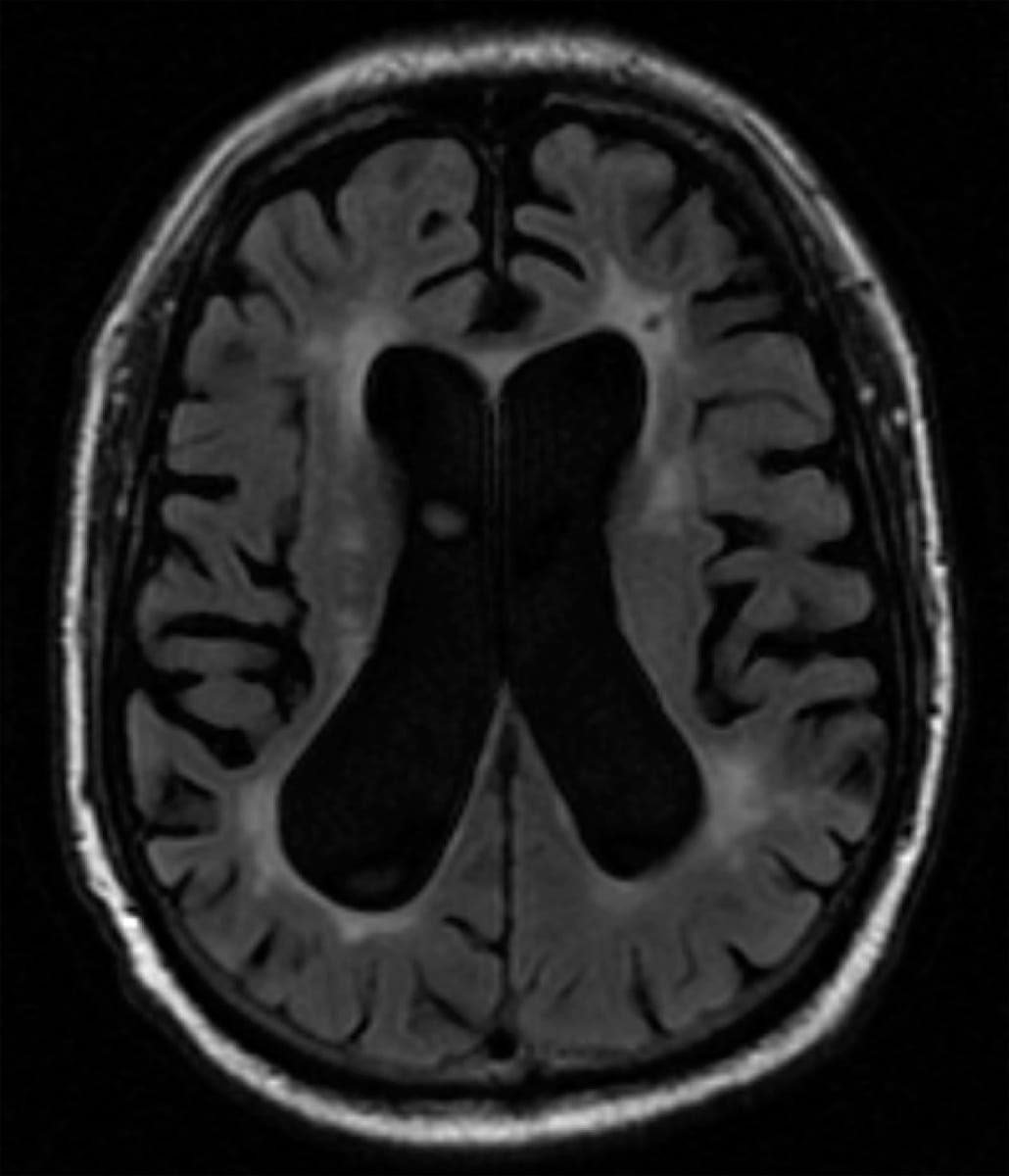
Vascular Dementia on brain MRI T2 FLAIR
Vascular Dementia
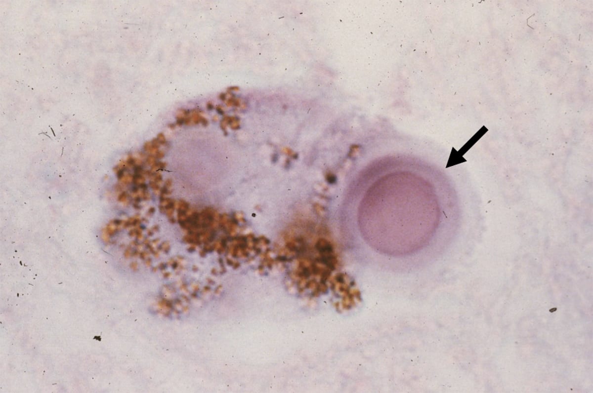
A synuclein lew body highpower
Lewy Body
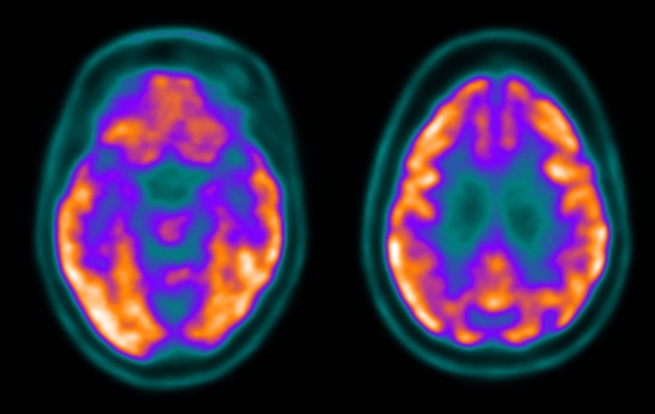
PET scan FTD
Frontotemporal Dementia
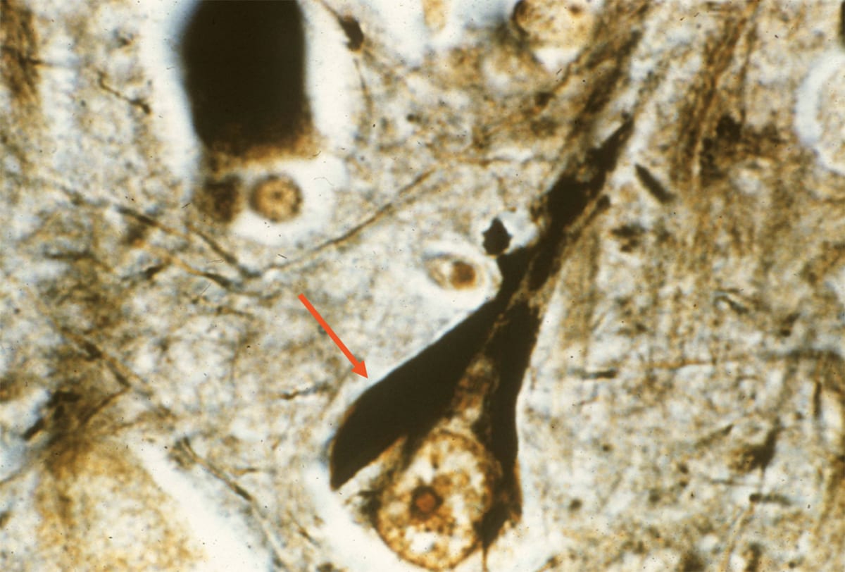
Neurofibrillary tangle silver stain alzheimer
Alzheimer's Disease
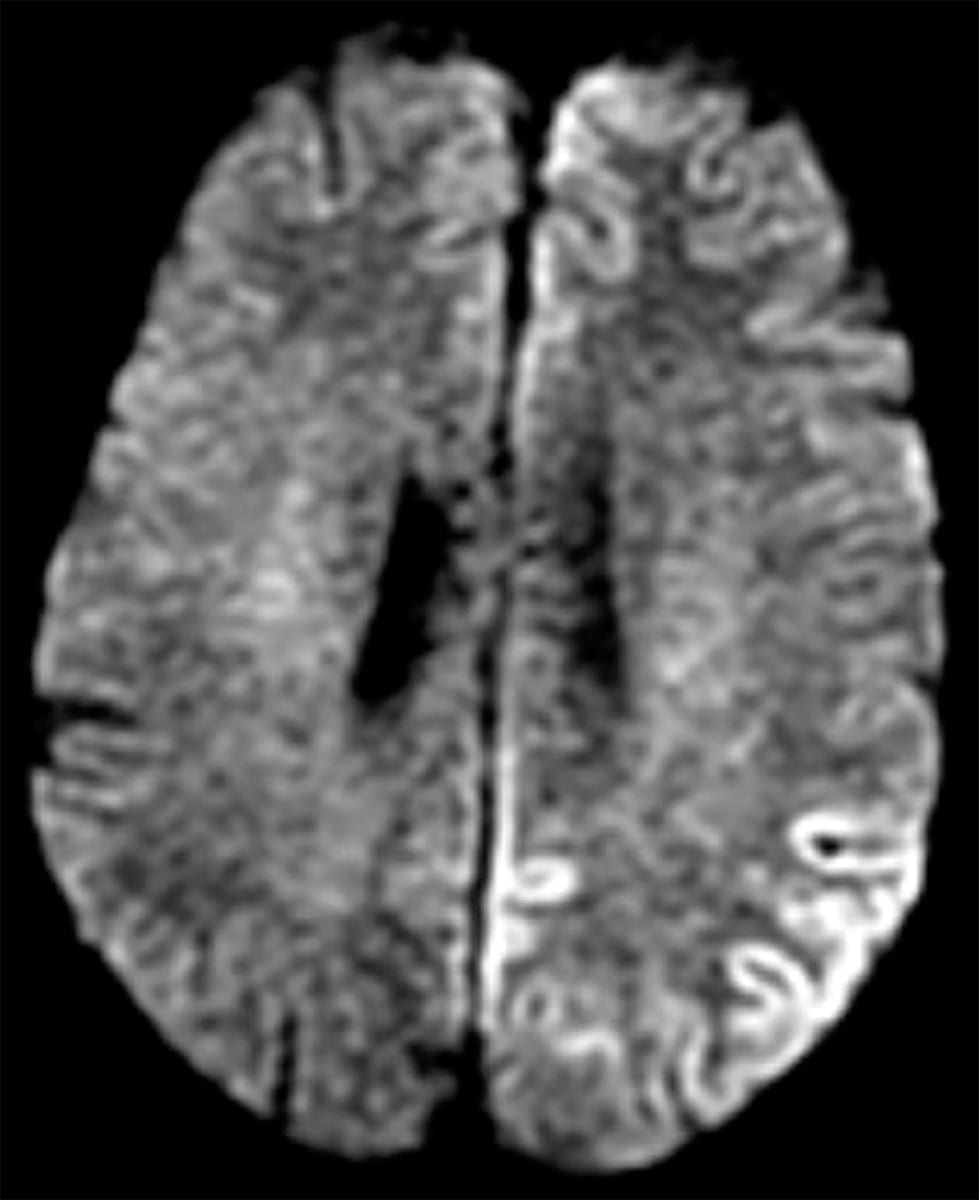
Creutzfeldt-Jakob Disease (CJD)
Cortical ribboning on axial brain MRI, DWI sequence.